← Reference

Fracture Classification Atlas

Pattern families first — eponyms become far less random once you see the logic behind them.

Study order: 1Pattern map → 2Regional retrieval → 3Decision trees → 4Image & case practice

Audition presentation formula

"This is a [classification name] [type/grade] fracture because [branch point finding], which matters because [clinical consequence]."
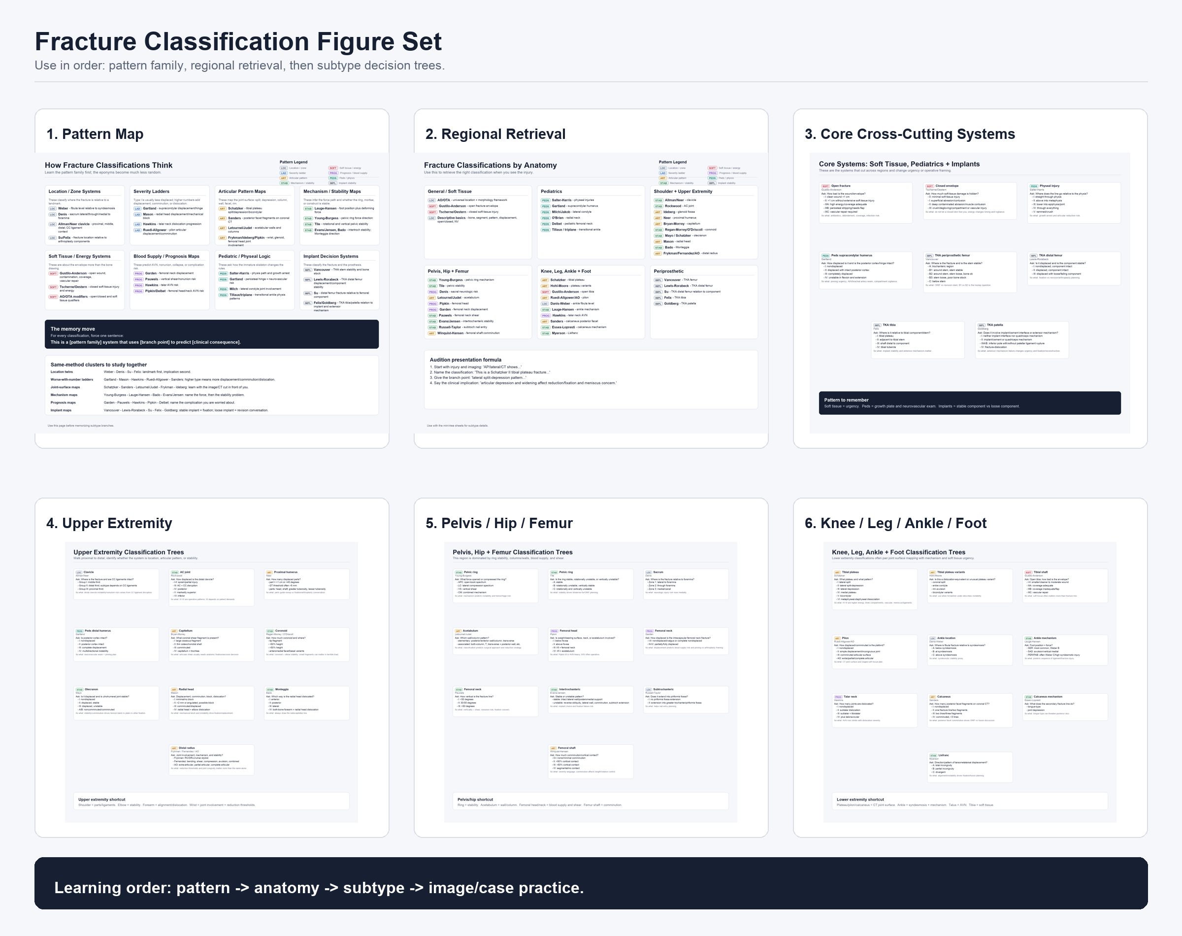
Figure set overview · Fig 00
Fracture Classification Figure Set

The complete six-panel index. Start here to see all six figure types at once and understand how the set is organized before drilling into individual panels.

LOC LAD ART STAB SOFT PROG PEDS IMPL
Six-panel index of the Fracture Classification Figure Set showing all study surfaces
Download

Study prompts

Learning orderWhat are the four steps in the recommended study sequence?
Pattern familiesName all eight pattern family types before looking.
MethodFor every classification: force one sentence using the pattern — [family] system that uses [branch point] to predict [clinical consequence].
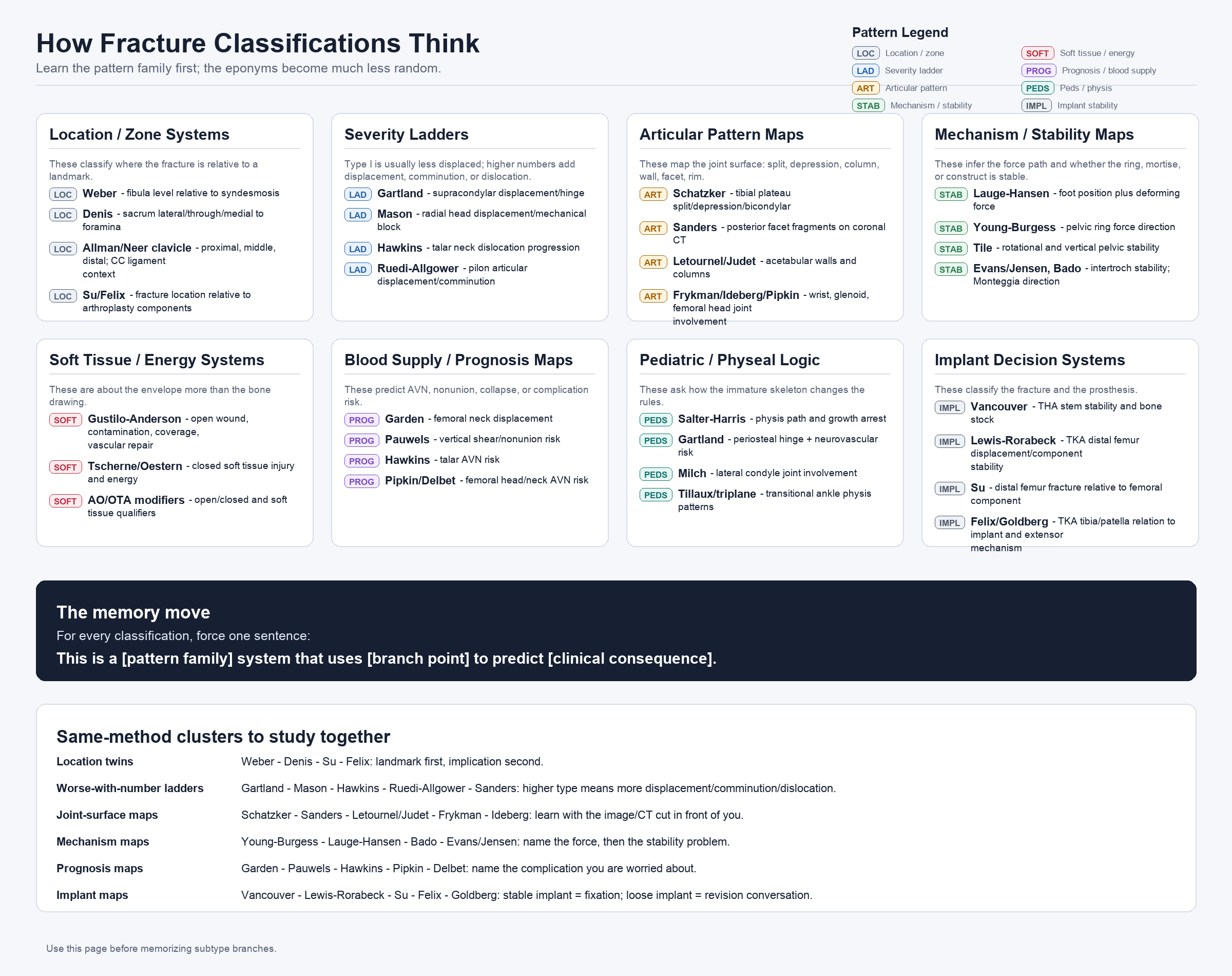
Pattern map · Fig 01
How Fracture Classifications Think

Eight pattern families with same-method clusters. Use this figure before memorizing any subtype branches — the eponyms become far less random once you understand which logic family each system belongs to.

LOC · Location/zone LAD · Severity ladder ART · Articular pattern STAB · Mechanism/stability SOFT · Soft tissue/energy PROG · Prognosis/blood supply PEDS · Peds/physis IMPL · Implant stability
How Fracture Classifications Think: eight pattern family boxes with same-method study clusters and memory move formula
Download

Study prompts

Pattern familyWhich pattern family does Gartland belong to? Weber? Garden? Letournel/Judet?
Memory moveSay the memory sentence for Schatzker, Hawkins, and Young-Burgess without looking.
Same-method clustersName the four severity ladders. Name the four prognosis maps.
So whatWhat does a higher number usually predict in a severity-ladder system?

Audition presentation formula — regional retrieval

1. Start with imaging: "AP/lateral/CT shows…"
2. Name the classification: "This is a Schatzker II tibial plateau fracture…"
3. Give the branch point: "lateral split-depression pattern…"
4. State the clinical implication: "articular depression and widening affect reduction/fixation and meniscus concern."
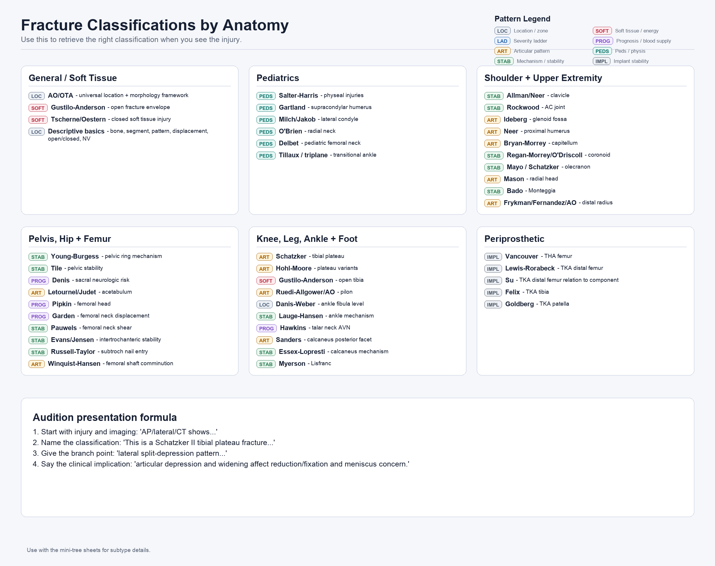
Regional retrieval map · Fig 02
Fracture Classifications by Anatomy

Use this when you know the injury location but need to recall which classification applies. Grouped by region with pattern-family tags. The audition presentation formula is at the bottom.

General/Soft Tissue Pediatrics Shoulder + UE Pelvis, Hip + Femur Knee, Leg, Ankle + Foot Periprosthetic
Fracture Classifications by Anatomy: regional grid grouping all major classification systems by injury location with pattern-family badges
Download

Study prompts

RetrievalCover the figure. Name all periprosthetic classifications. Then uncover and check.
Branch pointWhich ankle classifications complement each other — location vs mechanism?
Pattern familyWhich Pelvis/Hip classifications are PROG (prognosis/blood supply)?
PresentationPick any injury on this figure. Say the full audition formula out loud.
Core cross-cutting systems · Fig 03
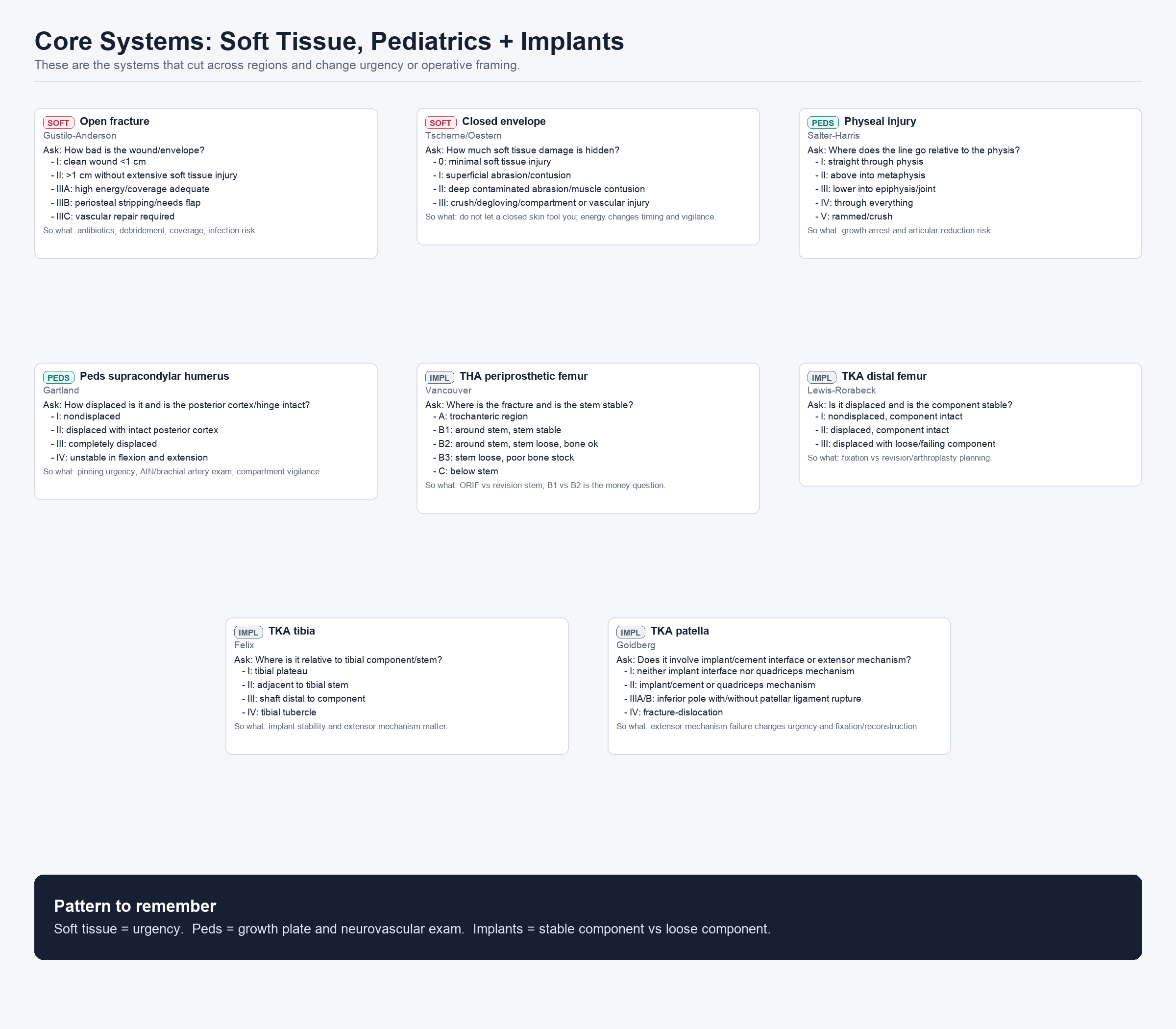
Core Systems: Soft Tissue, Pediatrics + Implants

The systems that cut across all regions and change urgency or operative framing. Gustilo-Anderson, Tscherne, Salter-Harris, Gartland, and the four periprosthetic systems (Vancouver, Lewis & Rorabeck, Felix, Goldberg).

SOFT · Open + closed envelope PEDS · Physeal + supracondylar IMPL · THA + TKA periprosthetic
Core Systems figure: Gustilo-Anderson, Tscherne/Oestern, Salter-Harris, Gartland, Vancouver, Lewis-Roabeck, Su, Felix, and Goldberg classification trees
Download

Study prompts

Pattern to remember"Soft tissue = urgency. Peds = growth plate and neurovascular exam. Implants = stable vs loose component." Can you explain why each part is true?
GartlandWhat makes Gartland III different from II? What exam is mandatory?
VancouverB1 vs B2 — what single finding changes the plan?
Gustilo-AndersonWhat distinguishes IIIA from IIIB? Why does that distinction matter operatively?

How to use decision trees

Each tree answers one diagnostic question. Walk the branches in order. Ask → find the finding → name the grade → state the "so what." If you can say the "so what" without the tree, you own the classification.
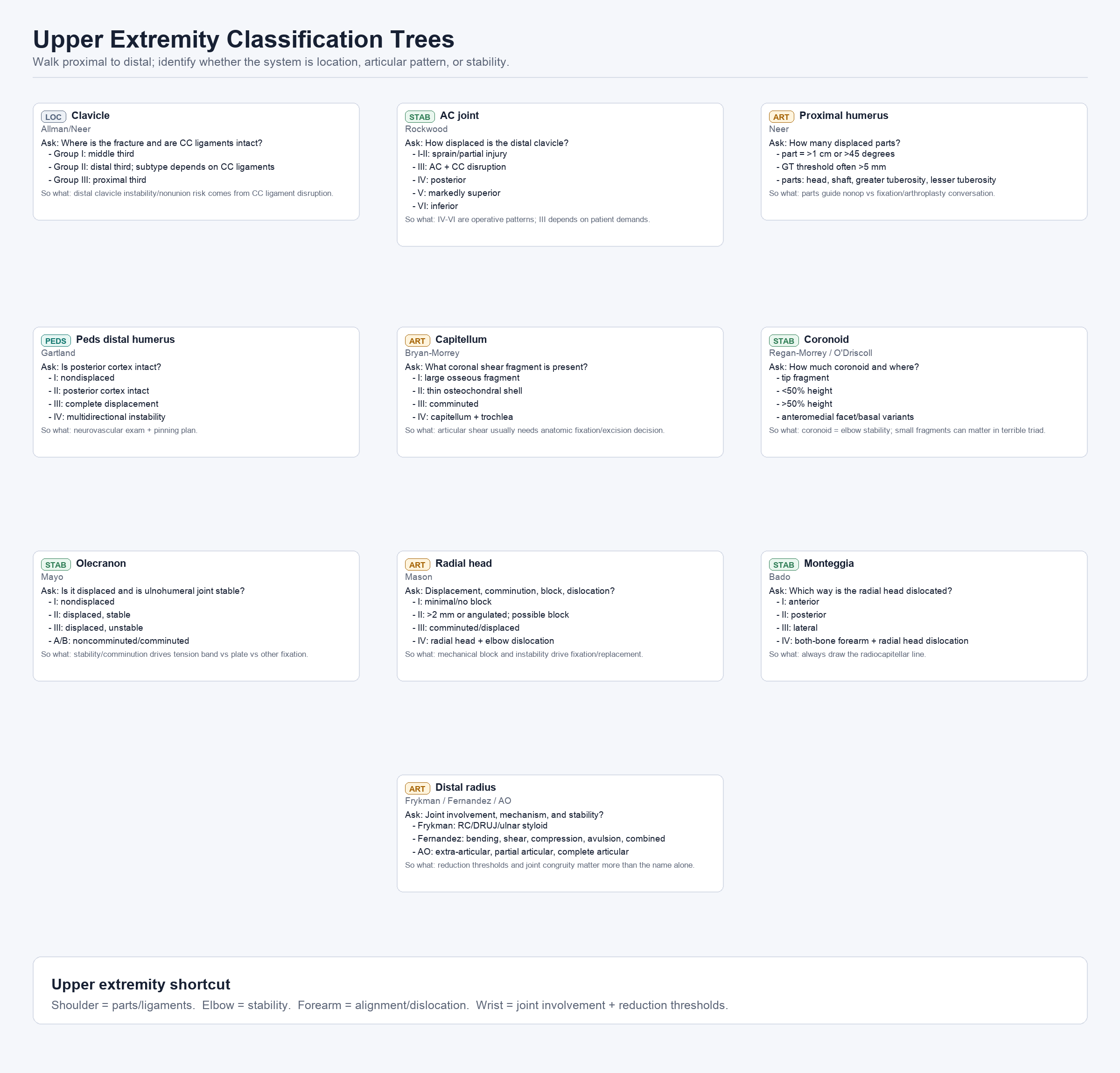
Decision trees · Fig 04
Upper Extremity Classification Trees

Clavicle (Allman/Neer), AC joint (Rockwood), proximal humerus (Neer), peds distal humerus (Gartland), capitellum (Bryan-Morrey), coronoid (Regan-Morrey/O'Driscoll), olecranon (Mayo), radial head (Mason), Monteggia (Bado), distal radius (Frykman/Fernandez/AO).

LOC STAB ART PEDS
Upper Extremity Classification Trees: decision trees for clavicle, AC joint, proximal humerus, elbow, forearm, and wrist fractures
Download

Study prompts

UE shortcut"Shoulder = parts/ligaments. Elbow = stability. Forearm = alignment/dislocation. Wrist = joint involvement + reduction thresholds." Test each one.
NeerWhat two thresholds define a displaced fragment? Name the four parts.
MasonWhat finding moves a radial head fracture from Mason II to Mason III?
MonteggiaWhat must you always check on any forearm fracture X-ray? Why?
Decision trees · Fig 05
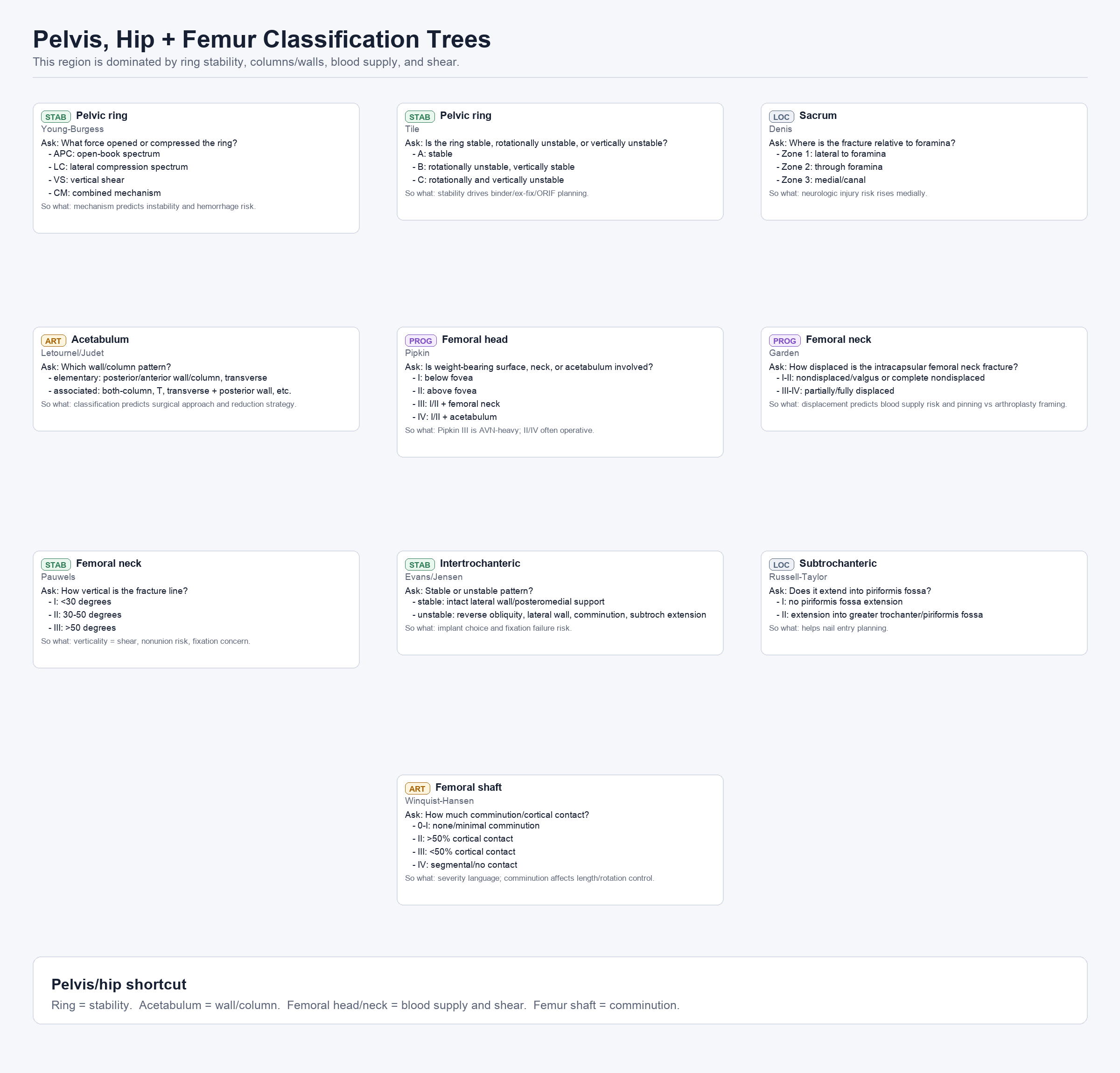
Pelvis, Hip + Femur Classification Trees

Young-Burgess, Tile (pelvic ring), Denis (sacrum), Letournel/Judet (acetabulum), Pipkin (femoral head), Garden (femoral neck displacement), Pauwels (femoral neck shear), Evans/Jensen (intertrochanteric), Russell-Taylor (subtrochanteric), Winquist-Hansen (femoral shaft).

STAB · Ring stability LOC · Sacrum, subtroch ART · Acetabulum, femoral shaft PROG · Femoral head/neck
Pelvis, Hip and Femur Classification Trees: decision trees for pelvic ring, sacrum, acetabulum, femoral head/neck, intertrochanteric, subtrochanteric, and femoral shaft fractures
Download

Study prompts

Pelvis/hip shortcut"Ring = stability. Acetabulum = wall/column. Femoral head/neck = blood supply and shear. Shaft = comminution." Explain each in one sentence.
GardenGarden III vs IV — what is the clinical difference and why does it matter?
PipkinWhy is Pipkin III AVN-heavy? What does Pipkin IV add?
Young-BurgessWhich pattern predicts the highest hemorrhage risk?
Decision trees · Fig 06
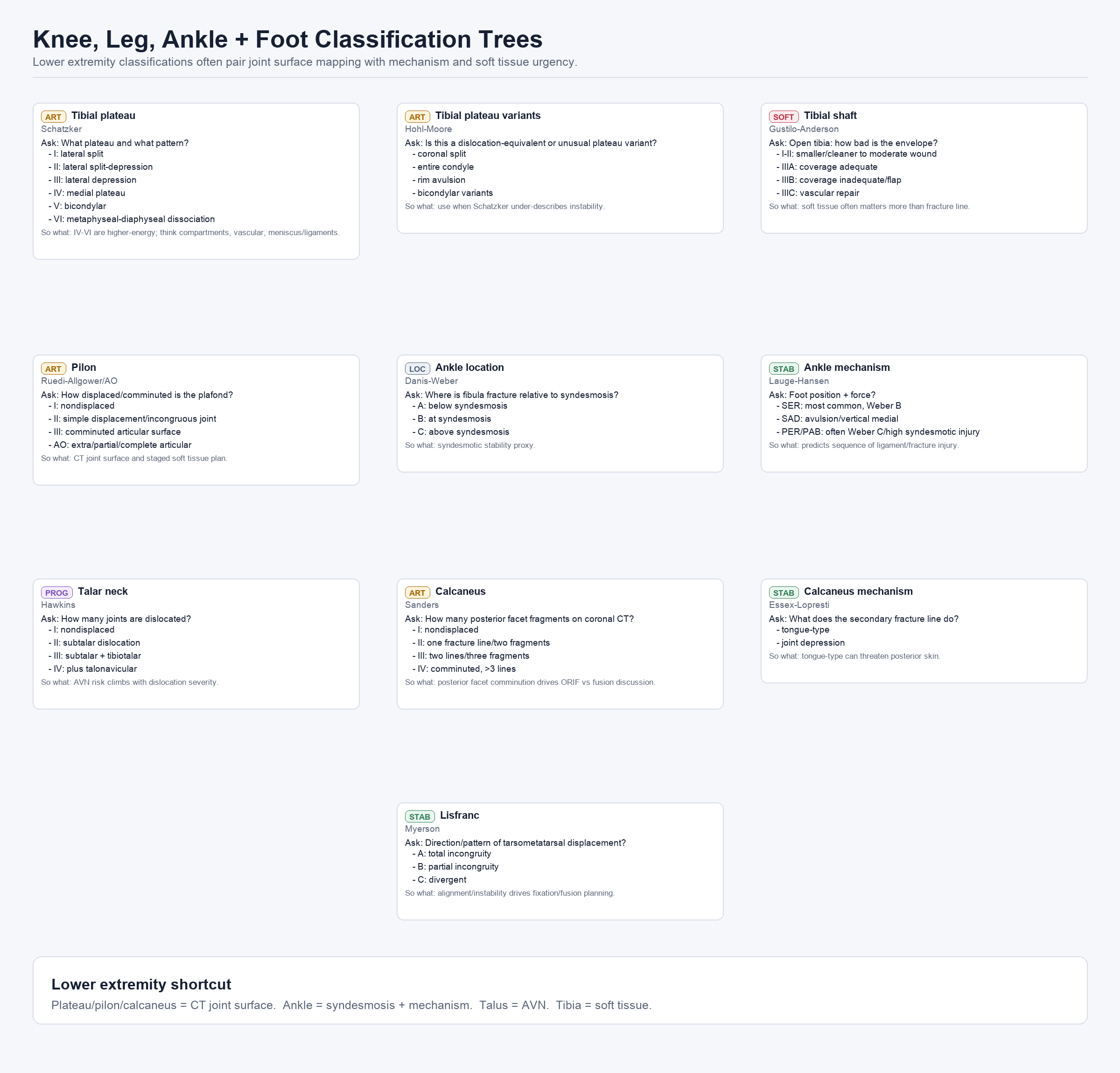
Knee, Leg, Ankle + Foot Classification Trees

Schatzker (tibial plateau), Hohl-Moore (plateau variants), Gustilo-Anderson (open tibia), Ruedi-Allgower/AO (pilon), Danis-Weber (ankle location), Lauge-Hansen (ankle mechanism), Hawkins (talar neck AVN), Sanders (calcaneus), Essex-Lopresti (calcaneus mechanism), Myerson (Lisfranc).

ART · Tibial plateau, pilon, calcaneus LOC · Ankle fibula level STAB · Ankle mechanism, Lisfranc PROG · Talar neck AVN SOFT · Open tibia
Knee, Leg, Ankle and Foot Classification Trees: decision trees for tibial plateau, pilon, ankle, talus, calcaneus, and Lisfranc fractures
Download

Study prompts

LE shortcut"Plateau/pilon/calcaneus = CT joint surface. Ankle = syndesmosis + mechanism. Talus = AVN. Tibia = soft tissue." Explain the reasoning.
SchatzkerWhich types are higher-energy? What associated structures should you worry about?
HawkinsHow many joints are dislocated in Hawkins IV? What is the AVN risk?
Danis-Weber vs Lauge-HansenWhat question does each system answer? When would you use one vs the other on rounds?